Integument Histology
The integument is comprised of the skin, sweat glands, sebaceous glands, nails and hair.
THE SKIN IS THE LARGEST ORGAN OF THE HUMAN BODY AND IS COMPOSED OF THE EPIDERMIS AND DERMIS LAYERS
The skin is an external coat to underlying bodily tissues and serves several important functions:
1) Protection: protects against abrasive injuries, bacterial invasion, and water loss
2) Body temperature regulation: blood vessels of the skin will constrict or dilate to aid in thermoregulation in response to cold or heat, respectively
3) Receptive of the external environment: skin receptors allow for perception of pain, temperature, and pressure
4) Excretion: Glands of the skin secrete fluids to assist in thermoregulation
5) Absorption: Ultraviolet rays are absorbed through the skin and are necessary for Vitamin D synthesis
I. Epidermis/Dermis/Hypodermis
The epidermis is derived embryologically from ectoderm and is compromised of a keratinized stratified squamous epithelium. The epidermis interdigitates closely with the dermis layer, which is located deep to the epidermis. A structure known as the rete apparatus is responsible for the attachment between the epidermis and the dermis and consists of dermal ridges (papillae) and epidermal ridges, which belong to the dermis and epidermis layers, respectively. Compared to the epidermis, the dermis is derived from mesoderm and is composed of dense irregular connective tissue responsible for strength, mechanical support, and increasing the thickness of the skin. An even deeper layer, the hypodermis, is a fatty layer known as the panniculus adiposus that is influenced by in caloric intake (ex: obesity) or environment (ex: cold climate). The hypodermis is not considered a layer of the skin but rather a layer of superficial fascia immediately deep to the dermis.
The skin varies by body region such that some locations have thick or thin skin (eyelids) and can be even devoid of hair (palms of the hands, soles of feet).
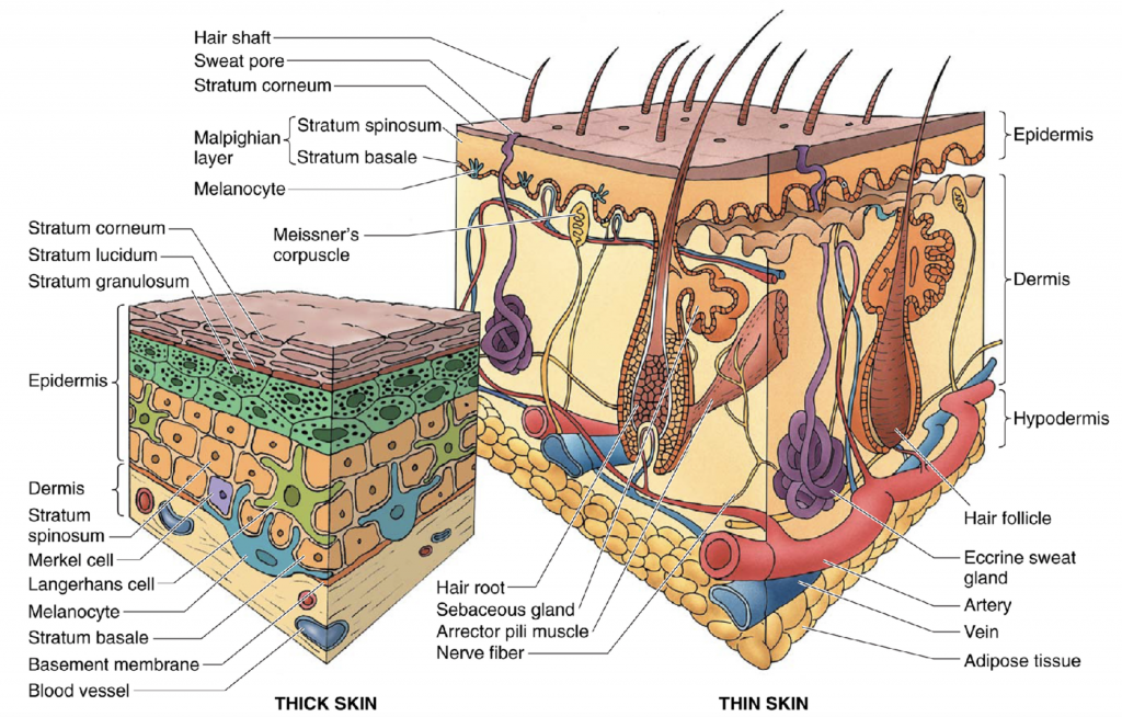
Image: Gartner & Hiatt (2007). Color Textbook of Histology: Saunders Elsevier, p. 328
a) Epidermis:
The epidermis consists of 4 distinct cell layers, or 5 distinct cells layers if the skin is derived from thick skin. The epidermis consists of stratified squamous epithelium with each layer displaying unique characteristics. The keratinized stratified squamous epithelium is composed of keratinocytes with three other cells types found amongst them: melanocytes, Langerhans cells, and Merkel cells
1) Stratum basale (AKA: stratum germinativum): These cells typical appear small cuboidal to low columnar with large nuclei and are mitotically active (mitotic figures often present) due to their function in keratinocyte renewal. The cells contain less cytoplasm than the stratum spinosum and are more basophilic. The basal cells of the stratum basale contain melanin at their apical surfaces transferred in from neighboring melanocytes. Also found interspersed within this layer are Merkel cells, which are associated with sensory nerve endings. The basal cells (stem cells) are anchored to a basement membrane by hemidesmosomes and to adjacent basal cells and keratinocytes by desmosomes.
2) Stratum spinosum (“Prickle cells”): The cells of the stratum spinosum contain keratinocytes, which display an elaborate system of cytoplasmic extensions which are interconnected to adjacent processes from neighboring keratinocytes by desmosomes. This cell layer is also known as “Prickle cell” layer due to the cytoplasmic extensions emanating from the keratinocytes. The keratinocytes appear polyhedral but become more flattened near the surface of this layer.
3) Stratum granulosum: The stratum granulosum is composed of 3 to 5 layers of flattened keratinocytes. This layer is aptly named due to the presence of non-membrane-bound keratohyalin granules. The keratohyaline granules are irregularly-shaped and stain intensely basophilic in histological preparations. The stratum granulosum also contains lamellar bodies (membrane-coating granules) which are released by exocytosis into the extracellular space. The lamellar bodies (membrane-coating granules) are lipid-rich and bathe the extracellular surfaces creating an impenetrable water barrier as the stratum granulosum becomes the stratum corneum.
4) Stratum corneum: The cells of the stratum corneum are the most differentiated cells of the epidermis but are anucleate and flattened. The stratum corneum is bathed in the lipid-rich membrane-coating granules released from subjacent stratum granulosum cells and form an impenetrable water barrier. The stratum corneum varies in thickness depending on the forces subjected upon the region.
5) Stratum lucidum (Thick skin only): The stratum lucidum is considered a subdivision of the stratum corneum by some histologists and appears in thick skin. The cells stain poorly hence the name stratum lucidum, or clear layer.
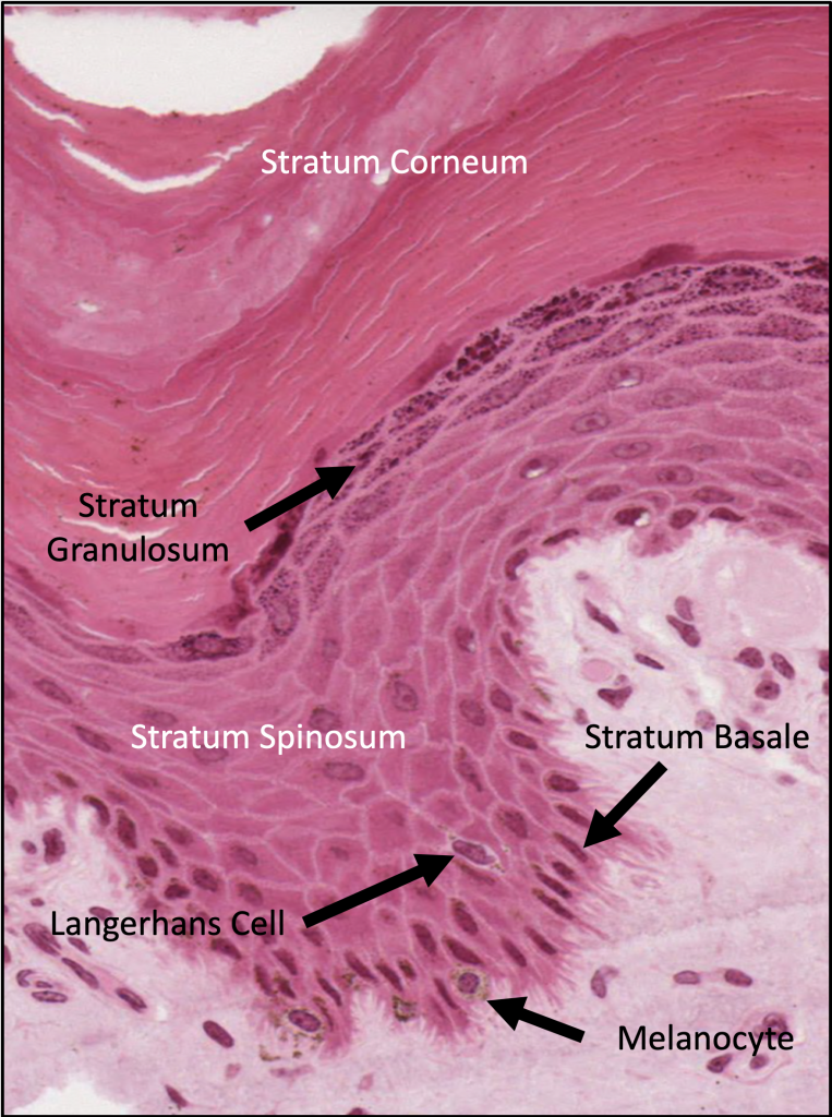
Image: Dr. Chapman – Virtual Microscopy Database (American Association of Anatomists)
Clinical correlation:
Basal Cell Carcinoma: The most common type of skin cancer is basal cell carcinoma. This type of cancer occurs when the epidermal cells of the stratum basale become malignant tumors due to long-term ultraviolet light exposure. The basal cells usually don’t metastasize and the tumors are treated through surgical removal.
Squamous Cell Carcinoma: The second most common type of skin cancer is squamous cell carcinoma. This type of cancer is characterized by highly atypical cells at all levels of the epidermis. Known for variable differentiation patterns from polygonal to rounded cells with or without orderly lobule arrangement and keratinization. Squamous cell carcinoma can become malignant and metastasize if the basement membrane becomes disrupted. Treatment with radiation, chemotherapy, or a more precise Moh micrographic procedure may be needed beyond simple surgical excision.
Malignant Melanoma: The most serious form of skin cancer is malignant melanoma due to its potential for metastasis. This type of cancer originates from melanocytes in the epidermis. Melanoma cells contain large nuclei with irregular contours and prominent eosinophilic nucleoli. Melanoma cells grow outwards in radial growth phase into the epidermis and dermis. During the vertical growth phase, an dark irregularly-shaped pigmented nodule appears on the skin with black, brown or red color variations. The melanoma cells may spread to regional lymph nodes as the cancer progresses.
b) Dermis:
The dermal-epidermal junction is not a linear boundary but rather an irregular boundary consisting of a series of protrusions from the dermis (dermal papillae) and epidermis (epidermal or rete ridges). Regions susceptible to greater mechanical forces have longer closely-packed dermal epidermal ridges. The dermal papillae create dermatoglyphics, which are genetically unique to each individual and form fingerprints.
The dermis is subdivided into two regions: Papillary Layer & Reticular Layer
i.) Papillary Layer:
The papillary layer is composed of loose connective tissue and thin elastic fibers. This layer is named for its location within the dermal papillae, which contains blood vessels and nerve endings at the dermal-epidermal junction.
ii.) Reticular Layer:
The reticular layer is composed of thick irregular bundles of type I collagen and coarse elastic fibers. The collagen and elastic fibers are aligned in parallel to the skin surface and form Langer’s lines, which are routinely incised in parallel during skin incisions to facilitate skin healing post-surgical procedures. Sweat glands, sebaceous glands, hair follicles, and arrector pili (smooth muscle attached to hair follicles) is found within the deep dermis. Several encapsulated mechanoreceptors are located in the deepest portions of the dermis: pacinian corpuscles (pressure and vibration), Ruffini corpuscles (tensile forces, most abundant in soles of feet), Krause end bulbs. The panniculus adiposus is found deep to the reticular layer.
II. Epidermal Cells
a) Keratinocytes
The immature keratinocytes contain a large amount of free ribosomes responsible for the synthesis of keratin granules, which accounts for the basophilia of these cells early in their development in the stratum basale. The keratin granules aggregate into keratin filaments (tonofilaments) and stain eosinophilic in the stratum spinosum. Keratinization takes between 2 and 6 hours as the cells progress from the stratum granulosum to the stratum corneum. Desquamation is controlled by pH-dependent proteases that breakdown the desmosomes of the keratinocytes in the stratum corneum.
b) Melanocytes
Melanocytes are derived from neural crest cells and migrate to the epidermis during development. The melanocytes contain processes that contact several keratinocytes in the stratum spinosum and collectively referred to as the “epidermal melanin unit”. Melanosomes are oval vesicles containing tyrosinase created by the RER of the melanocyte. Melanosomes are extruded into the extracellular space and phagocytksed by the keratinocytes of the stratum spinosum. The tyrosinase converts tyrosine into melanin and is transported to the supra nuclear region of the keratinocyte to protect the chromosomes from UV radiation. Over a couple of days, the melanin is eventually attacked and degraded by lysosomes within the keratinocyte, which provides an explanation for why light-skinned individuals eventually lose their tan, unless they have continued sun exposure.
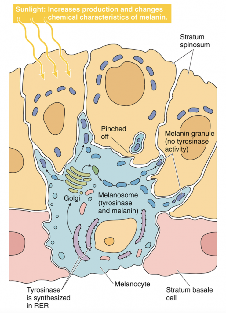
Image: Gartner & Hiatt (2007). Color Textbook of Histology: Saunders Elsevier, p. 334
The number of melanocytes are similar amongst light- and dark-skinned individuals; however, the difference in skin pigmentation is due to a greater rate of tyrosinase activity and larger size of melanosomes of dark-skinned individuals.
Clinical correlation:
Albinisim: absence of melanin production due to genetic defect in tyrosine synthesis. Melansomes containing the amino acid tyrosine are present within the melanocytes but they are not converted into melanin due to failure of tyrosinase synthesis.
Amino Acid Tyrosine –> converted by Tyrosinase (produced by RER of melanocyte) –> Melanin granule (within melanosomes)
c) Merkel cells
Merkel cells are derived from neural crest cells embryologically. These cells are found interspersed among the stratum basale and are attached to keratinocytes of the adjacent stratum spinosum by desmosomes. Myelinated sensory neurons closely approximate the Merkel cells forming Merkel cell-Neurite complexes, which suggest they act as mechanoreceptors, especially in the skin of the fingertips. There are several key histological features of the Merkel cell: indented/lobed nucleus, dense-cored granules in supranuclear region and processes.
d) Langerhans (Dentritic) cells
Langerhans cells are derived from the bone marrow and are considered part of the mononuclear phagocyte system, acting as an antigen presenting cell (APC). These cells are located in the stratum spinosum and are often described as dentritic cells due to their large cytoplasmic processes. Using light microscopy, Langerhans cells are characterized by a pale cytoplasm, dense nucleus and processes radiating from the cell body. In electron micrographs, the most unique feature of the Langerhans cells are membrane-bound Birbeck granules, which resemble the shape of Ping Pong paddles. The Birbeck granules assist in engulfing and degrading antigens phagocytosed by the Langerhan cells. The cells are mobile and will migrate to lymph nodes where they present epitopes to T-lymphocytes enabling an immune response.
III. Glands of the Skin
The glands of the skin include eccrine sweat glands, apocrine sweat glands and sebaceous glands.
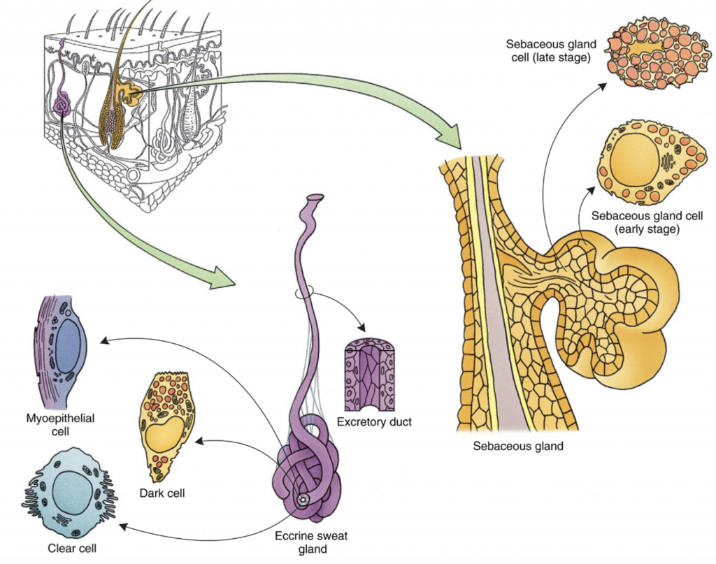
Image: Gartner & Hiatt (2007). Color Textbook of Histology: Saunders Elsevier, p. 337
a) Eccrine Sweat Glands (merocrine secretion)
Eccrine sweat glands are organs of thermoregulation and their secretions can range from 1L to 10L/day depending on physical activity levels and warm climates. Eccrine sweat glands are found in the deep dermis and are simple coiled tubular glands. Upon stimulation by the sympathetic nervous system, the eccrine glands secrete sweat through merocrine secretion in that their proteins are exocytosed into the lumen with no loss of cytoplasm. The excretory ducts of the gland reach the skin surface as a sweat pore.
SECRETORY PORTION: Eccrine secretory end pieces contain Dark cells and Clear cells:
Dark cells: dark cells appear as inverted cones and obscure the clear cells from touching the secretory lumen of the gland. Dark cells secrete a mucous-rich substance.
Clear cells: clear cells appear as cones with a large base attached to the basement membrane and narrow apical segment and is devoid of secretory granules. The cells secrete a watery mucous that mixes with dark cells secretions.
Myoepithelial cells: the eccrine secretory end piece is surrounded by myoepithelial cells which contain contractile filaments, actin and myosin, which expel fluid from the secretory portion into the duct of the gland.
DUCT PORTION: Eccrine secretory end pieces are continuous with the duct portion, which contains stratified cuboidal epithelium
Basal cell layer: consist of large, heterochromatic nuclei with abundant mitochondria
Luminal cell layer: consists of irregularly shaped nucleus with spare cytoplasm and few organelles
b) Apocrine Sweat Glands (Merocrine Secretion)
Apocrine sweat glands are limited to certain bodily regions including the armpit, areola and anal region. These glands lack typical ducts like those found in the eccrine sweat glands and empty into channel surrounding hair follicles. The apocrine sweat glands empty just superficial to the entry of the sebaceous glands (see below) along the hair follicle. The secretory cells of Apocrine sweat glands are simple cuboidal to low columnar. These glands are innervated by the sympathetic nervous system and are under influence of hormones, which explains that their development doesn’t occur until puberty.
c) Sebaceous Glands (Holocrine Secretion)
Sebaceous glands secrete sebum, which is an oily/wax-like substance consisting of cholesterol, triglycerides and cellular debris from the secretory cells. Functionally, the sebum maintains hair flexibility, skin texture and provides antimicrobial protection in addition to acting as a water barrier. Similar to apocrine sweat glands, the sebaceous glands release sebum into the upper third of the hair follicle, which nourishes the growing hair follicle.
SECRETORY PORTION: The sebaceous glands are lobular in structure with clusters of acini opening into short ducts. The acini consist of small peripherally-located basal cells with larger round cells. The large round cells are filled with lipid droplets and eventually become necrotic and the lipid and cellular components are released into the lumen (holocrine secretion)
DUCT PORTION: The short sebaceous gland ducts are lined by stratified squamous epithelium continuous with the follicular canal
Clinical correlation:
Acne – the sebaceous glands become clogged by cellular debris and sebum with concomitant bacterial infection produces a chronic inflammatory response surrounding the hair follicles
IV. Hair Follicles
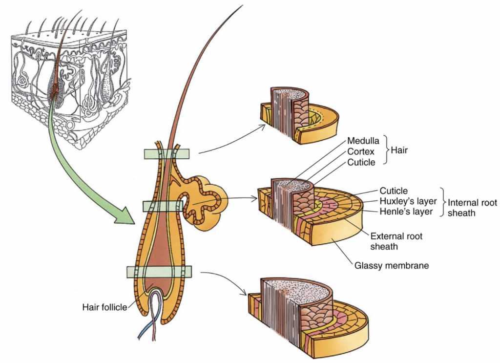
Image: Gartner & Hiatt (2007). Color Textbook of Histology: Saunders Elsevier, p. 341
Hair follicles are surrounded by dense fibrous connective tissue derived from the dermis layer. The glassy membrane (basement membrane) separates the dermis from the epithelium of the hair follicle itself. The hair root is indented with the dermal papilla projecting into the base of the root. The dermal papilla contains a blood supply to provide nutrients to the cells of the hair root. Basal cells, also known as matrix cells, proliferate and contribute to the formation of hair. Melanocytes scattered amongst the matrix cells contribute melanosomes, which contribute to hair pigmentation.
The matrix cells develop to form the internal root sheath:
Cuticle: squamous cells facing the hair shaft
Huxley’s layer: single or double layer of squamous cells that form the middle plate of the internal root sheath
Henle’s layer: an outer single layer of cuboidal cells (in contact with innermost layer of cells of the external root sheath).
The shaft of the hair is composed of three regions:
Medulla: large vacuolated cells that form the core of the hair shaft
Cortex: cells at the periphery of the medulla
Cuticle of the hair: cells at the periphery of the medulla form the cuticle of the hair
V. Integument Sensory Organs
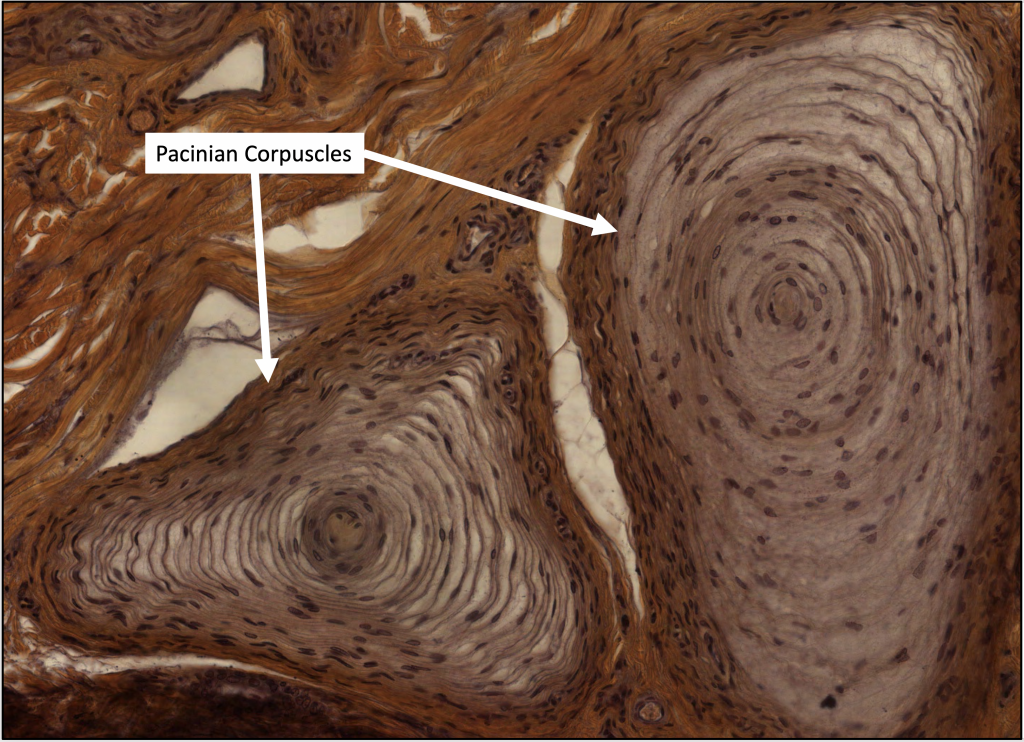
a) Pacinian Corpuscle
Pacinian corpuscles appear as large encapsulated ovoid lamellar structures found in the deep dermis or hypodermis. They frequently appear in bodily areas in which fine touch sensation is predominant such as in the fingertips. Pacinian corpuscles respond to pressure and vibration through displacement of the capsule lamellae which are filled with a lymph-like fluid. The displacement of the lamellae depolarizesof the axon found within.
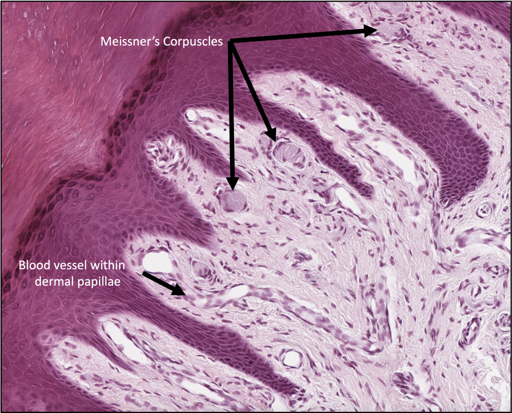
b) Meissner’s Corpuscle
Meissner’s corpuscle are touch receptors that respond primarily to low frequency stimuli in the papillary layer of hairless skin (lips, dorsal or ventral surfaces of the palm). The corpuscles appear as cylinders present in the dermal papillae beneath the basal lamina of the epidermis. Schwann cells found in the corpuscle form irregular flattened lamellae.
c) Ruffini’s Corpuscle
Ruffini’s corpuscles are encapsulated mechanoreceptors consisting of an elaborate nerve arborization that is sensitive to sustained or continuous mechanical stress such as stretch and torque. These mechanoreceptors are rapid adapting to skin stress.
Images derived from: Gartner & Hiatt (2007). Color Textbook of Histology: Saunders Elsevier, pp. 327-344