In short, we work on developing optical imaging technologies for biomedical applications! Our research intersects the multidisciplinary fields of biomedical imaging, optics, electrical and computer engineering, machine learning, and medicine. We aim to impact medical device and imaging technologies impacting patient diagnosis, treatment monitoring, and predicting disease progression.
Handheld Optical Coherence Tomography
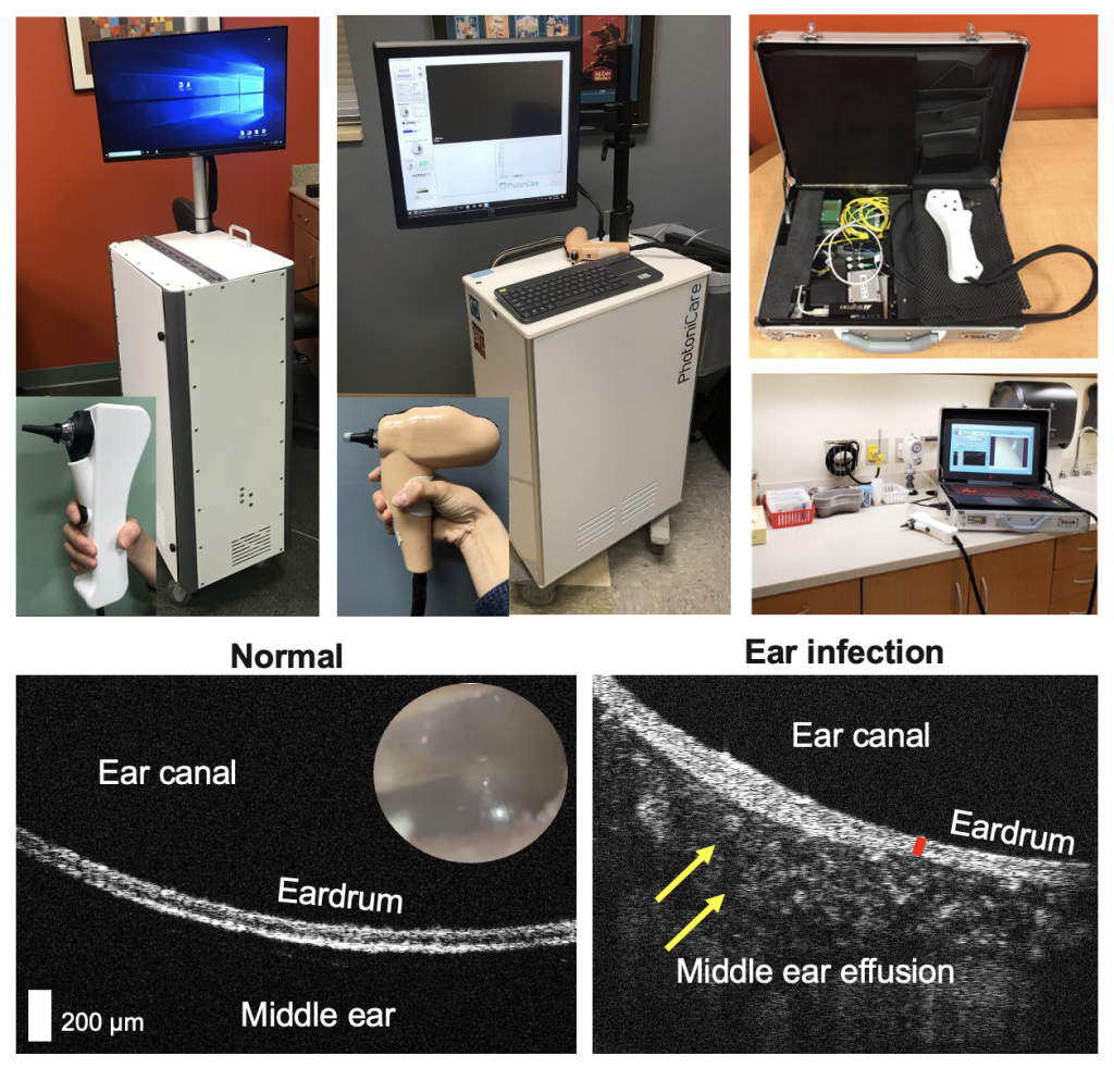
Most of our work focus on optical coherence tomography (OCT). You can think of it as ‘optical’ version of ultrasound imaging, where we can generate cross-sectional images inside tissue, but utilizing light. The standard OCT imaging contrast comes from the differences in refractive index of tissue layers or boundaries. One of the applications that we are working on is to non-invasively visualize the middle ear cavity, to study prevalent middle ear infections (otitis media). To facilitate biomedical research studies, we have developed different formfactors of handheld OCT devices, ranging from a portable cart to a briefcase OCT device.
We work towards developing low-cost, compact OCT devices while maintaining adequate system performance for versatile biomedical applications.
Ophthalmic Optical Coherence Tomography
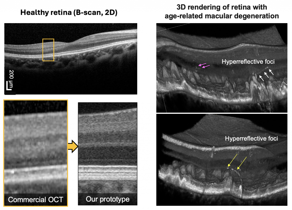
Ophthalmology is by far the most successful clinical application of optical coherence tomography (OCT), resulting in rapid clinical adoption since its invention in early 1990s. High axial resolution and sufficient imaging depth allowed efficient imaging of retina, through our lens of the eye as an optical component focusing the OCT beam. For the past decades, researchers have focused on improving OCT technologies, in terms of speed, resolution, size, and creating additional capability (e.g. OCT angiography, elastography, etc). Recent advancements in lasers and optoelectronics have enabled ultrahigh resolution and ultrahigh speed OCT instruments that are clinically feasible to use with patients.
We work towards developing clinically feasible, advanced OCT prototype instruments and analytical methods to better understand retinal diseases.
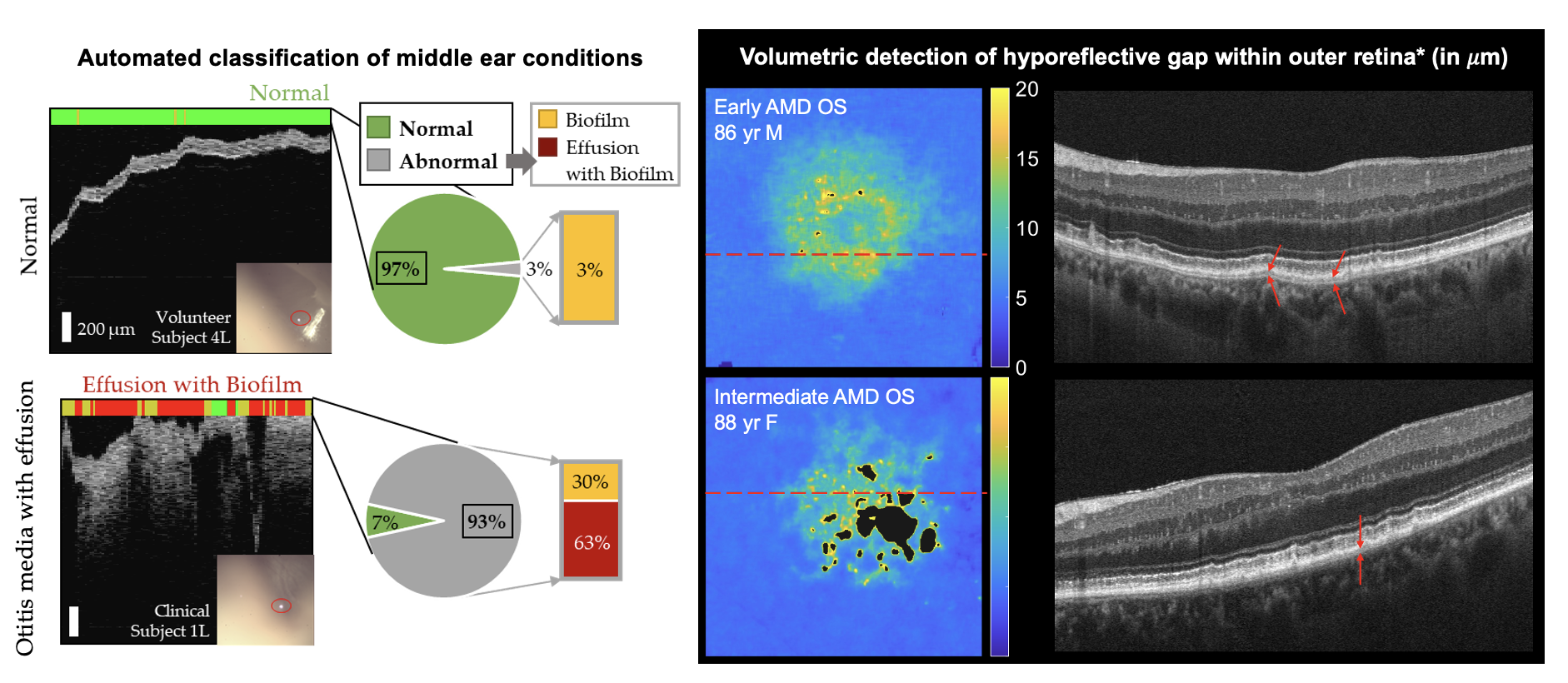
Machine Learning Driven Assessment of Biomarkers
We work with multidimensional images! This often includes 3D spatial coordinates (x, y, z), time (t), and sometimes additional imaging contrasts! Thus, we closely work with machine learning and computer vision methods for various tasks, including but not limited to classification, segmentation, and prediction. We investigate ‘feature of interest’ or potential imaging biomarkers in various diseases, such as age-related macular degeneration, diabetic retinopathy, and middle ear infections!
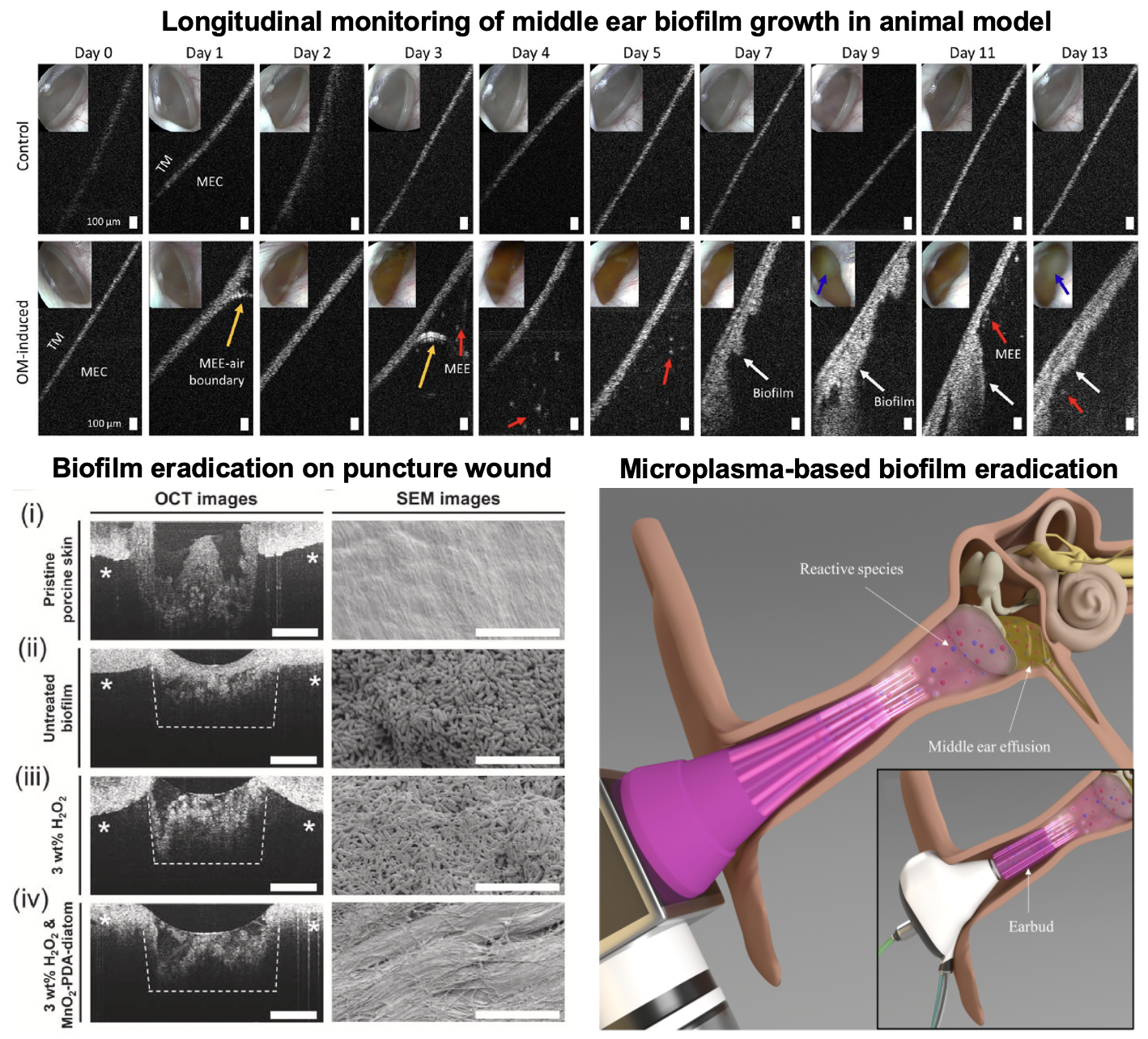
Non-destructive Bacterial Biofilm Characterization
We study bacterial biofilms, or more specifically, we develop imaging tools and analytical methods to help studying bacterial biofilms. Bacterial biofilms are accumulated or clustered bacteria adhered to a surface, and thus are more resistant to antibiotics and changes in the external environment compared to bacteria. Bacterial biofilm is ubiquitous in biomedicine, i.e. dental bacterial biofilm, biomaterials and implant, medical devices inserted to patients, water biofilm, and middle ear biofilm. We have utilized our imaging technologies to non-destructively study bacterial biofilm growth and eradication methods and efficacy via in vitro, lab-grown biofilms, as well as in animal models. Furthermore, we can also detect and assess middle ear biofilm adherent to the eardrum, inside pediatric patients with chronic ear infections.